Philomorph with a sense of adventure and lots of curiosity
Menu
Philomorph with a sense of adventure and lots of curiosity
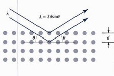
In my ’20s I fell in love with the methods by which we are able to discover and visualize the three-dimensional structures of molecules using X-Ray and neutron diffraction. Crystallography, at the intersection of chemistry, mathematics and computing, fit my academic and professional aspirations, my skills and my nature, like a glove.
My Ph.D was in X-Ray crystallography of small molecules; my post-doc in protein crystallography; and my research position at the Institut Laue Langevin in Grenoble, France, combined instrument development, software development, and service crystallography.
I worked on the crystal structures of organometallic complexes when “FGAS” – Professor F. Gordon A. Stone – was head of the inorganic chemistry department in the School of Chemistry at the University of Bristol, in the UK.
Prof. Stone wrote an autobiography published as one of a series by the American Chemical Society. The book is “Leaving No Stone Unturned : Pathways in Organometallic Chemistry“, F.Gordon A.Stone, American Chemical Society, Washington, DC 1993, from the series “Profiles, Pathways, and Dreams: Autobiographies of Eminent Chemists”, Jeffrey I.Seeman, Series Editor.
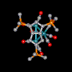
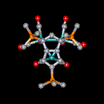
Illustrated are two isomeric forms of triruthenium carbonyl clusters with face- and edge-bridging pentalene ligands from publication A2.
A11 Dimolydenum Complexes containing Bridging Silyl-substituted Acetylenes: Crystal and Molecular Structure of [Mo2(CO)4(μ-Me3SiC2SiMe3)(η-C5H5)2].
J.A.Beck, S.A.R.Knox, R.F.D.Stansfield, F.G.A.Stone, M.J.Winter and P.Woodward,
Journal of the Chemical Society, Dalton Transactions, (1982) 195-200.
A10 Dimolydenum Complexes derived from Cyclo-octa-1,5-diene and Penta-1,3-diene. Crystal and Molecular Structure of [Mo2(CO)3(μ-C8H10)(η-C5H5)2].
M.Griffiths, S.A.R.Knox, R.F.D.Stansfield, F.G.A.Stone, M.J.Winter and P.Woodward,
Journal of the Chemical Society, Dalton Transactions, (1982) 159-165.
A9 Dimolybdenum Complexes derived from Cyclo-octatetraene. Crystal and Molecular Structures of [Mo2(CO)2(η-C5H5)2(C8H8)] (Two Isomers) and of [Mo2(CO)4(η-C5H5)2(η3,η'3-C16H16)].
R.Goddard, S.A.R.Knox, R.F.D.Stansfield, F.G.A.Stone, M.J.Winter and P.Woodward,
Journal of the Chemical Society, Dalton Transactions, (1982) 147-158.
A8 Reactions of Cyclic 1,3-Dienes with Ethyne co-ordinated at a Dimetal Centre. Crystal Structure of [Mo2(CO)2(μ-η2,η'2-C10H10)(η-C5H5)2].
S.A.R.Knox, R.F.D.Stansfield, F.G.A.Stone, M.J.Winter and P.Woodward,
Journal of the Chemical Society, Dalton Transactions, (1982) 167-172.
A7 Reduction-Oxidation Properties of Organotransition-metal Complexes. Part 12. Formation of Carbon-Carbon Bonds via the Oxidative Dimerisation of [Fe(CO)3(η4-C8H8)] and the Reduction of [Fe2(CO)6(η5:η'5-C16H16)]2+; X-Ray Crystal Structures of [Fe2(CO)4{P(OPh)3}2(η5:η'5-C16H16)][PF6]2 and [Fe(CO)3(η4-C16H16)].
N.G.Connelly, R.L.Kelly, M.D.Kitchen, R.M.Mills, R.F.D.Stansfield, M.W.Whiteley, S.M.Whiting and P.Woodward,
Journal of the Chemical Society, Dalton Transactions, (1981) 1317-1326.
A6 The Sequential Linking of Alkynes at a Dimolybdenum Centre. Crystal Structures of [Mo2{μ-(MeO2CC2CO2Me)(HC2H)(MeO2CC2CO2Me)2}(η-C5H5)2].2CH2Cl2 and [Mo2{μ-(MeO2CC2CO2Me)4}(η-C5H5)2].
A.M.Boileau, A.G.Orpen, R.F.D.Stansfield and P.Woodward,
Journal of the Chemical Society, Dalton Transactions, (1982) 187-193.
A5 The Sequential Linking of Alkynes at Dichromium and Dimolybdenum Centres; X-Ray Crystal Structure of [Cr2(CO)(μ-C4Ph4)(η-C5H5)2].
S.A.R.Knox, R.F.D.Stansfield, F.G.A.Stone, M.J.Winter and P.Woodward,
Journal of the Chemical Society, Dalton Transactions, (1982) 173-185.
A4 Alkyne Complexes of Platinum. Part 4. Stepwise Formation of Di- and Tri-platinum Complexes with Bridging Alkyne Ligands; Crystal Structure of [Pt3{μ-(η2-PhC2Ph)}2(PEt3)4].
N.M.Boag, M.Green, J.A.K.Howard, J.L.Spencer, R.F.D.Stansfield, M.D.O.Thomas, F.G.A.Stone and P.Woodward,
Journal of the Chemical Society, Dalton Transactions, (1980) 2182-2190.
A3 η2 Complexes of Cyclic Polyolefins: Crystal Structure of [Mn(CO)2(η2-C8H8)(η-C5H5)].
I.B.Benson, S.A.R.Knox, R.F.D.Stansfield and P.Woodward,
Journal of the Chemical Society, Dalton Transactions, (1981) 51-55.
A2 Pentalene Complexes of Ruthenium: Two Isomeric forms of Triruthenium Carbonyl Clusters with Face- and Edge-bridging Pentalene Ligands: Crystal Structures of Two Isomers of [Ru3(CO)8{C8H3(SiMe3)3-1,3,5}].
J.A.K.Howard, R.F.D.Stansfield and P.Woodward,
Journal of the Chemical Society, Dalton Transactions, (1979) 1812-1818.
A1 Crystal Structures of Two Closely Related Compounds of Molybdenum-(II) and -(IV): Carbonylcyclopentadienylhexafluorobut-2-yne(penta-fluorophenylthio)molybdenum and Cyclopentadienyl(hexafluorobut-2-yne)oxo(pentafluorophenylthio)molybdenum.
J.A.K.Howard, R.F.D.Stansfield and P.Woodward,
Journal of the Chemical Society, Dalton Transactions, (1976) 246-250.
Post-doctoral work in the Department of Biochemistry (now the School of Biosciences) at Sheffield University, UK, under the direction of Prof. Pauline Harrison, on the structure of horse spleen apoferritin (ferritin without its iron core).
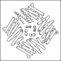
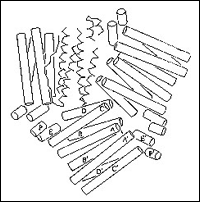
a) Eight apoferritin subunits form a channel around each end of three axes of 4-fold symmetry, in a molecule formed of 24 identical subunits. The crystal space group is F432 !
b) The second diagram shows six apoferritin subunits around one of the four axes of 3-fold symmetry. Both these diagrams come from the letter to Nature, B8 below.
C2 The Apoferritin Shell and its Assembly: Some 3-Dimensional Views.
P.E.Bourne, P.M.Harrison, D.W.Rice, J.M.A.Smith and R.F.D.Stansfield,
Proceedings of the Fifth International Conference on Proteins of Iron Storage and Transport, U.C. San Diego, U.S.A., August 23-27, 1981.
C1 The Structure and Function of Ferritin: Past Progress and Future Promise.
P.E.Bourne, P.M.Harrison, W.G.Lewis, D.W.Rice, J.M.A.Smith and R.F.D.Stansfield, Proceedings of the Fifth International Conference on Proteins of Iron Storage and Transport, U.C. San Diego, U.S.A., 23-27 August, 1981.
B8 Helix packing and subunit conformation in horse spleen apoferritin.
G.A.Clegg, R.F.D.Stansfield, P.E.Bourne and P.M.Harrison,
Nature, 288 (1980) 298-300.
B7 The structure and heavy-metal-ion-binding sites of horse spleen apoferritin.
G.A.Clegg, R.F.D.Stansfield, P.E.Bourne and P.M.Harrison,
Biochemical Society Transactions, 8(5) (1980) 654-655.
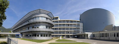
Work done at the Institute Laue Langevin, Grenoble, France as a “Physicien” in the Diffraction group in the 1980’s.
I was co-responsible for the development of the Neutron Diffractometer D19 equipped with a Position Sensitive Detector for protein crystallography.
The work, all of which I loved, included developing the instrument, hosting scientists who visited to perform diffraction experiments, and pursuing original research.
A18 X-Ray and Neutron Diffraction Study of the Crystal Structure of MnS2.
T.Chattopadhyay, H.G. von Schnering, R.F.D.Stansfield and G.J.McIntyre,
Zeitschrift für Kristallographie, 199 (1992) 13-24.
A17 Binding of Dimethyl Sulfoxide to Lysozyme in Crystals, Studied with Neutron Diffraction.
M.S. Lehmann and R.F.D. Stansfield,
Biochemistry, 28 (1989) 7028-7033.
A15 A High-Resolution Neutron-Diffraction Study of the Effects of Deuteration on the Crystal Structure of KH2PO4.
Z.Tun, R.J.Nelmes, W.H.Kuhs and R.F.D.Stansfield,
Journal of Physics C: Solid State Physics, 21 (1988) 245-258.
A13 Hydrido-silyl-komplexe VII. Strukturchemische und 29Si-NMR-Spectroskopische Unterschungen an C5R'5(CO)(L)Mn(H)SiR3. Einfluss der Substituenten R und R' sowie des Liganden L auf die Mn, H, Si-dreizentrenbindung.
U.Schubert, G.Scholz, J.Mueller, K.Ackermann, B.Worle und R.F.D.Stansfield,
Journal of Organometallic Chemistry, 306 (1986) 303-326.
A12 The Effect upon the Hydrogen Atoms of Bonding an Allyl Group to a Transition Metal. A Theoretical Investigation and an Experimental Determination using Neutron Diffraction of the Structure of Bis(η3-allyl)nickel.
R.Goddard, C.Kruger, F.Mark, R.F.D.Stansfield and X.Zhang,
Organometallics, 3 (1985) 285-290.
B9 Structural determination by neutron diffraction of oriented deuterated trans-polyacetylene.
G.Pepy, L.Rosta, B.Francois, C.Mathis, G.J.McIntyre and R.F.D.Stansfield,
Annales de Physique, (Colloque C1), 11 (1986) 81-82.
A16 Integration of Single Crystal Reflections Using Area Multidetectors.
C.Wilkinson, H.W.Khamis, R.F.D.Stansfield and G.J.McIntyre,
Journal of Applied Crystallography, 21 (1988) 471-478.
A14 A General Lorentz Correction for Single-Crystal Diffractometers.
G.J.McIntyre and R.F.D.Stansfield,
Acta Crystallographica, A44 (1988) 257-262.
C8 Neutron Area Diffractometry and Spatial Deconvolution Techniques.
R.F.D.Stansfield and C.Wilkinson, Proceedings of the Daresbury Study Weekend 23-24 January, 1987, Computational Aspects of Protein Crystal Data Analysis, DL/SCI/R25, SERC Daresbury Laboratory, 1987, Eds. J.R.Helliwell, P.A.Machin and M.Z.Papiz.
C7 Possible Use of OCCAM and Transputers in PSD Data Treatment.
S.A.Wilson and R.F.D.Stansfield, Proceedings of an International Workshop held at Grenoble, France, 4-6 November, 1985, Journal de Physique, (Colloque 5), 47 (1986) 193-197.
C6 On-line Evaluation of PSD Diffraction Data.
R.F.D.Stansfield and G.J.McIntyre, Proceedings of an IAEA (International Atomic Energy Authority) Conference held at Julich, Federal Republic of Germany, 14-18 January, 1985, Neutron Scattering in the 'nineties, IAEA, Vienna, 1985, 191-198.
C5 D19A and B: Design and Construction of a 4-Circle Neutron Diffractometer with Two-Dimensional PSD's.
M.Thomas, R.F.D.Stansfield, M.Berneron, A.Filhol, G.Greenwood, J.Jacobe, D.Feltin and S.A.Mason, in Position Sensitive Detection of Thermal Neutrons, Eds. P.Convert and J.B.Forsyth, Academic Press, London, 1983, 344-350.
C4 Tests of the Quality of Data using a "Fly's-Eye" Neutron PSD.
R.F.D.Stansfield, M.Thomas, S.A.Mason, R.J.Nelmes, J.E.Tibballs and W.L.Zhong,
in Position Sensitive Detection of Thermal Neutrons, Eds. P.Convert and J.B.Forsyth,
Academic Press, London, 1983, 365-371.
C3 Multidetector Development: Tests with a Phthalocyanine Crystal.
R.F.D.Stansfield,
AIP (American Institut of Physics) Conference Proceedings No. 89, Neutron Scattering - 1981, (Argonne National Laboratory), Ed. J.Faber Jnr., New York, 1982, 196-198.
Copyright © 2024 | Rob Stansfield | All rights reserved